


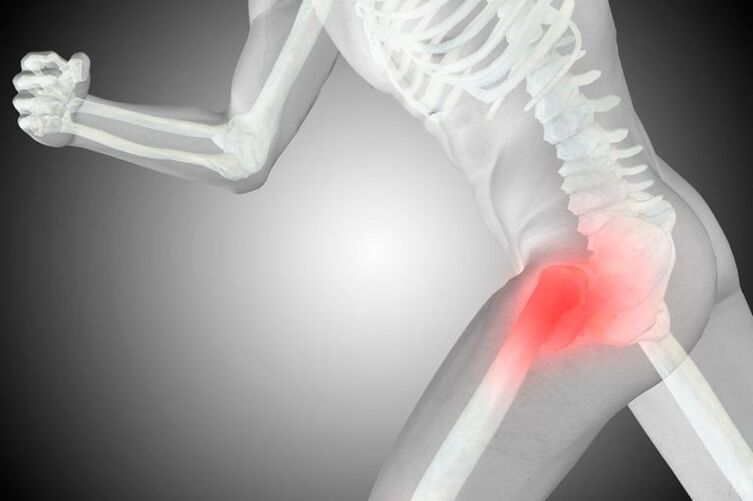
Coxarthrosis is a common degenerative-dystrophic disease of the hip joint, where, due to age-related changes or other factors, there is gradual destruction of the articular joint of the femoral head and pelvic acetabulum. It is accompanied by pain and limitation of the amplitude of movement of varying severity, which depends on the stage of development. And if at the initial stage it is possible to overcome coxarthrosis with conservative methods, then at the 3rd stage it is possible to save the condition and maintain the working capacity of the hip joint, that is, to avoid deformity, only by performing an operation.
It belongs to the number of arthrosis and may be accompanied by the development of the same process in other joints, and this pathology accounts for about 12% of all diseases of the musculoskeletal system. But the term "coxarthrosis" can only be used to describe degenerative-dystrophic changes in the hip joint.
What is coxarthrosis
Coxarthrosis is a complex pathology of one or both hip joints, in which the cartilaginous layer covering the femoral head and acetabulum is destroyed, which leads to a decrease in the size of the joint space. As the disease progresses, the appearance of bone surface deformation and the formation of bone growths on them, called osteophytes, are observed.
Coxarthrosis is the second most common disease in the musculoskeletal system. More often, only gonarthrosis is diagnosed, i. e. degenerative-dystrophic changes in the knee joint. However, the probability of disability in coxarthrosis is higher.
The entire hip joint is enclosed in a specific case, called the articular capsule. It has a so-called synovial membrane, which produces synovial fluid. This fluid is important for the proper functioning of the joint, as it not only lubricates the hyaline cartilage, but is also a source of nutrients for it.
Normally, cartilage is constantly worn out and quickly restored due to the continuous regeneration process, which is carried out with the help of substances that enter it from the synovial fluid. But with injuries or age-related changes, the rate of the regeneration process decreases, which leads to the gradual wear of hyaline cartilage and the development of coxarthrosis.
This is due to changes in the amount of synovial fluid produced and its composition. Under the influence of adverse factors, it becomes thicker and is produced in smaller quantities. As a result, the synovial fluid can no longer provide the hyaline cartilage with all the necessary substances in the right amount, which leads to dehydration and rapid thinning. Gradually, the strength and elasticity of the cartilage decreases, areas of delamination of the fibers that make it up, cracks form in it, and its thickness also decreases. These changes can be seen during instrumental diagnostic methods, in particular, the narrowing of the joint space draws attention to itself.
Narrowing of the joint space leads to increased friction between the bony structures that make up the hip joint and increased pressure on the already degrading hyaline cartilage. This creates more damage to it, which affects the function of the joint and the condition of the person, because the deformed area prevents the femoral head from sliding easily in the acetabulum. As a result, there are symptoms of coxarthrosis.
If left untreated, the pathological changes become worse, and the hyaline cartilage wears away. After that, in some areas, it disappears completely, which leads to bone exposure and a sharp increase in the load on the joints. Since when moving in the acetabulum, the femoral head rubs directly against the bone, this causes the appearance of severe pain and sharp limitation of movement. In this case, the pressure of bone structures on each other leads to the formation of bone growth on their surface.
Osteophytes that form can have sharp edges that can injure the muscles and ligaments surrounding the hip joint. This leads to the appearance of strong pain both directly in the joint area and in the groin, buttocks and thighs. As a result, the patient saves the injured leg, reduces pressure on it and tries to avoid making unnecessary movements with it. This causes the development of muscle atrophy, which worsens mobility disorders and eventually leads to lameness.
Cause
There are many reasons for the development of coxarthrosis, although in rare cases it occurs against the background of the absence of any prerequisites. In this case, they talk about the presence of primary or idiopathic coxarthrosis. In most cases, secondary coxarthrosis is diagnosed, which is a logical consequence of some disease or change in the state of the musculoskeletal system. It can be provoked by:
- hip joint injuries of various nature, including fractures, dislocations, bruises, sprains or torn ligaments;
- hard physical labor, professional sports, especially weightlifting, parachuting, jumping sports;
- sedentary lifestyle;
- overweight, which significantly increases the load on the hip joint;
- the focus of chronic infection in the body;
- congenital defects of the hip joint, such as dysplasia or dislocation;
- metabolic pathology and endocrine disorders, especially gout, diabetes mellitus, especially in the decompensated form;
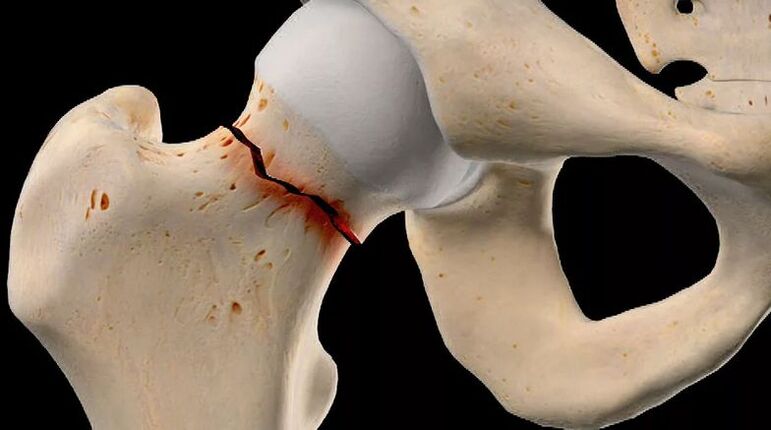
- aseptic necrosis of the femoral head, which may be the result of a fracture of the femoral neck, especially during conservative treatment;
- inflammatory diseases of the joints, including rheumatoid arthritis, bursitis, tendinitis;
- spinal disease;
- genetic predisposition;
- the presence of bad habits, especially smoking.
However, the main cause of coxarthrosis is still inevitable age-related changes, and the presence of the above factors only increases the likelihood and speed of its development.
Symptoms of coxarthrosis
The disease is characterized by a gradual development with a systematic increase in the intensity of symptoms. Therefore, in the early stages, it can be asymptomatic or only occasionally cause concern to the patient, but subsequently the condition of the hip joint worsens, which leads to an increase in the severity of the symptoms of coxarthrosis until unbearable pain and significant mobility limitations. .
So, degenerative-dystrophic changes in the hip joint are accompanied by:
- Pain of varying intensity, initially arising after physical exercise or walking and subsiding after rest. Gradually, the severity of the pain syndrome increases, it appears more often, lasts longer and the period between the moment of applying a load on the joint and the appearance of pain decreases. Then, the pain is present almost constantly, including at rest, and becomes unbearable. Increased pain at any stage of disease development during hypothermia and lifting heavy objects is characteristic.
- Restrictions on the mobility of the hip joint, which is initially indicated by small difficulties in the implementation of rotational movements of the legs. Over time, morning stiffness appears, which disappears after the patient "disperses. "This may be accompanied by the appearance of edema in the hip joint. As the disease progresses, mobility restrictions become more noticeable and persistent, that is, they do not disappear after warming up. Patients notice a decrease in the amplitude of leg movements, and subsequently completely lose the ability to perform certain movements.
- Cracks in the hip joint, which appear when walking or doing physical work, especially when doing extensions. It becomes the result of the friction of bare bone structures against each other, which is accompanied by a sudden increase in pain.
- Thigh muscle spasms, which lead to radiating pain in the thigh. This may be the result of the attachment of various intra-articular disorders, including nerve compression, excessive stretching of the ligaments around the joint, as well as the development of synovitis, that is, inflammation of the synovial membrane and accumulation of inflammation. drainage in the hip joint cavity.
- Lameness, which is initially the result of the patient's unconscious desire to reduce the load on the diseased joint and transfer the weight to the healthy leg to avoid the appearance or intensity of pain, and then the development of muscle contractures. The latter phenomenon already occurs in the final stage of coxarthrosis and leads to the fact that the patient cannot fully straighten the leg and, moreover, maintain it in this position. Therefore, the lower limb with the affected hip joint is always in a slightly bent position, which creates lameness.
- Reduction in leg length, which mainly occurs with severe degenerative-dystrophic changes in the hip joint, is accompanied not only by narrowing of the joint space, but also by flattening of the femoral head, muscle atrophy. As a result, the diseased leg becomes shorter than the healthy one by 1 or more centimeters.
Coxarthrosis can affect both one hip joint, and both at the same time. But if in the first case the symptoms of the disease will be observed only on one side, then in the second they will not only be bilateral, but will also differ in intensity. It depends on the degree of destruction of each hip joint.
Stage of coxarthrosis
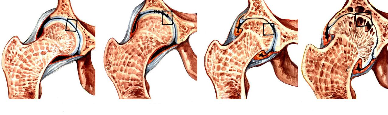
The nature of the manifestation of the disease depends on the stage of its development. In total, there are 3 degrees of coxarthrosis, where the first is considered the easiest. In the early stages of degenerative-dystrophic changes in the hip joint, only episodic pain can be observed. As a rule, this happens after vigorous physical exercise, playing sports or a long walk. Therefore, patients usually do not pay attention to them, associate them with fatigue and consider them normal age-related changes. In this regard, stage 1 coxarthrosis is diagnosed only in isolated cases, which usually occur during an examination for other reasons.
As the disease progresses, the symptoms intensify and already with coxarthrosis of the 2nd degree they feel themselves. The stage of development of this pathology is characterized by a narrowing of the joint space by 50%, as well as the appearance of signs of deformation of the femoral head with its displacement.
With the further development of the pathology, the joint space becomes narrower and with the 3rd degree of coxarthrosis it is almost completely absent. This is already accompanied by the formation of various osteophytes. At the advanced stage of the disease, the pain becomes not only strong, but unbearable and often occurs even in a state of complete rest, including at night. Because the hip joint is severely deformed, the elements can impinge on the nerves that pass through it, which leads to pain that radiates to the groin, back, as well as the thigh and even the lower leg. This also creates an inability to move independently without the use of aids, such as crutches or crutches.
Coxarthrosis of the 3rd degree is a direct indication for surgical treatment. If the operation is not performed in time, the femoral head will unite with the surface of the acetabulum with osteophytes. This will lead to the shortening of the legs, the complete absence of the possibility of free movement, because the joints will lose their mobility completely, that is, deformity.
Diagnostics
If signs of coxarthrosis occur, it is recommended to contact an orthopedic specialist as soon as possible. At first, the doctor will interview the patient and find out the nature of the complaint, and then proceed with the examination and the performance of functional tests, comparing the length of the legs. As a rule, the data obtained are sufficient to speak with a high degree of confidence about the presence of coxarthrosis.
But since such a clinical picture can accompany several other diseases of the hip joint, both inflammatory and non-inflammatory, instrumental diagnostic methods are needed. With their help, a specialist will be able not only to confirm the presence of coxarthrosis, to distinguish it from radicular syndrome caused by spinal pathology, but also to correctly assess the level of its development, which means choosing the most effective treatment tactics. .
Today for the diagnosis of coxarthrosis are used:
- X-ray of the hip joint - the resulting image allows you to detect signs of destructive changes, the presence of osteophytes, the nature of the deformation of the bone structure and measure the thickness of the joint space.
- CT is a more modern method for diagnosing bone pathology, providing clearer data than x-ray, but more expensive. Therefore, CT is prescribed in controversial cases, when it is necessary to clarify the diagnosis and the degree of destruction of the hip joint.
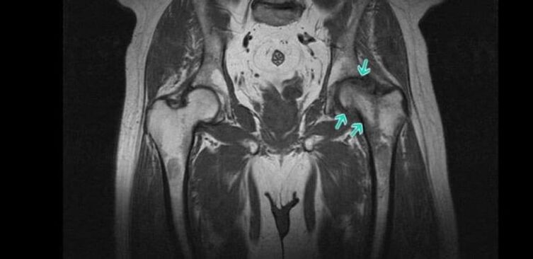
- MRI is a very informative method for examining the joint, providing the maximum amount of information about the state of the joint and all its structures, especially hyaline cartilage, ligaments, and blood supply characteristics.
Patients are prescribed several laboratory tests, including KLA, OAM, rheumatic tests, biochemical blood tests, etc.
Conservative treatment of coxarthrosis

When diagnosing coxarthrosis 1 or 2 degrees, treatment is carried out with conservative methods. For each patient, they are selected individually, taking into account the detected comorbidities. Therefore, it is often necessary to consult not only orthopedic specialists, but also doctors of other specialties who will choose the necessary treatment to combat concomitant diseases.
As part of the treatment of coxarthrosis, patients are prescribed:
- drug therapy;
- exercise therapy;
- physiotherapy.
It is mandatory for all patients to take measures to eliminate the effects of factors that increase the load on the legs and contribute to the development of degenerative changes in the hip joint. This includes adjusting your diet and increasing your physical activity level if you are overweight. If the patient is frequently exposed to excessive physical exertion, it is recommended to change the type of activity or reduce the intensity of the exercise, if the load is due to sports. In some cases, it is recommended to use special bandages and orthoses that will improve the hip joint and unload it during physical exercise.
Medical therapy
As part of drug treatment, the patient is individually selected drugs, taking into account existing concomitant diseases. As a rule, drugs of the following pharmacological groups are indicated for coxarthrosis:
- NSAIDs - a wide group of drugs that show analgesic and anti-inflammatory effects (available in various dosage forms, including tablets, capsules, gels, creams, injection solutions, which allow you to choose the most effective and convenient form of application);
- corticosteroids - drugs that have a strong anti-inflammatory effect, but due to the high risk of side effects, especially when using the oral form, they are only prescribed for short courses in the form of injections;
- muscle relaxants - drugs that help reduce muscle tone, which allows you to effectively deal with muscle spasms, often observed in coxarthrosis;
- chondroprotectors - a group of drugs that contain components used by the body for the regeneration of cartilage tissue;
- preparations that improve microcirculation - help improve soft tissue nutrition and activate metabolic processes in the affected area;
- Vitamin B - indicated for nerve conduction disorders caused by nerve compression by altered hip joint components.
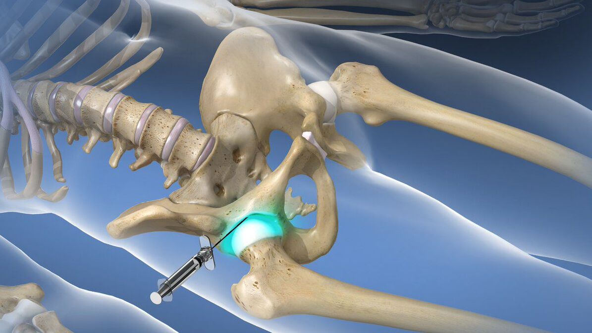
If coxarthrosis causes acute pain attacks, which cannot be stopped with the help of prescribed NSAIDs, intra-articular or periarticular blockade is recommended for the patient. Its essence lies in the introduction directly into the hip joint cavity of an anesthetic solution in combination with corticosteroids. This will allow you to quickly eliminate pain and reduce the inflammatory process. But restrictions can only be done by qualified health workers in a specially prepared room. Doing such a procedure at home is not indicated.
exercise therapy
When diagnosing coxarthrosis, regular exercise therapy is mandatory. In the same way as drug therapy, a set of exercise therapy exercises for each patient is selected individually, taking into account the degree of destruction of the hip joint, the level of physical development of the patient, the nature of concomitant diseases (special attention). paid to cardiovascular pathology).
Thanks to daily exercise therapy, you can:
- reduce the severity of pain;
- increase the mobility of the hip joint;
- reduce the risk of muscle atrophy;
- eliminate thigh muscle spasms;
- activates blood circulation and thereby improves the nutrition of the affected joints.
All exercises should be done smoothly, avoiding sudden movements and jerks. But if pain occurs during exercise therapy, you should definitely contact your doctor to correct the selected complex or carry out a re-diagnosis to exclude the development of the disease and the need for surgery.
Physiotherapy
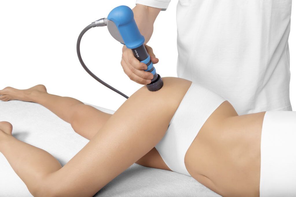
Comprehensive treatment of coxarthrosis involves a course of physiotherapy procedures that have anti-inflammatory, analgesic, decongestant and tonic effects on the body. Therefore, often patients are prescribed 10-15 procedures:
- ultrasound therapy;
- electrophoresis;
- UVT;
- magnetotherapy;
- laser therapy, etc.
Recently, plasmolifting has been increasingly used as part of the conservative treatment of coxarthrosis, which can significantly increase the speed of regeneration of hyaline cartilage. The essence of the procedure is the introduction into the cavity of the hip joint of purified blood plasma, obtained by centrifugation from the patient's own blood.
Surgery for coxarthrosis
If the patient is diagnosed with coxarthrosis of the 3rd degree, he is indicated for surgical intervention, because conservative methods in such cases are already powerless. Unfortunately, this kind of situation is very common today, because a large number of patients seek medical help when they can no longer tolerate pain or have serious mobility restrictions that prevent their ability to work and move freely.
Timely surgical intervention can completely eliminate this disorder and restore the patient's ability to move normally, significantly improving his quality of life. Indications for its implementation are:
- significant decrease in joint space by more than 80%;
- the presence of severe pain in the hip joint, which cannot be eliminated;
- obvious mobility impairment;
- femoral neck fracture.
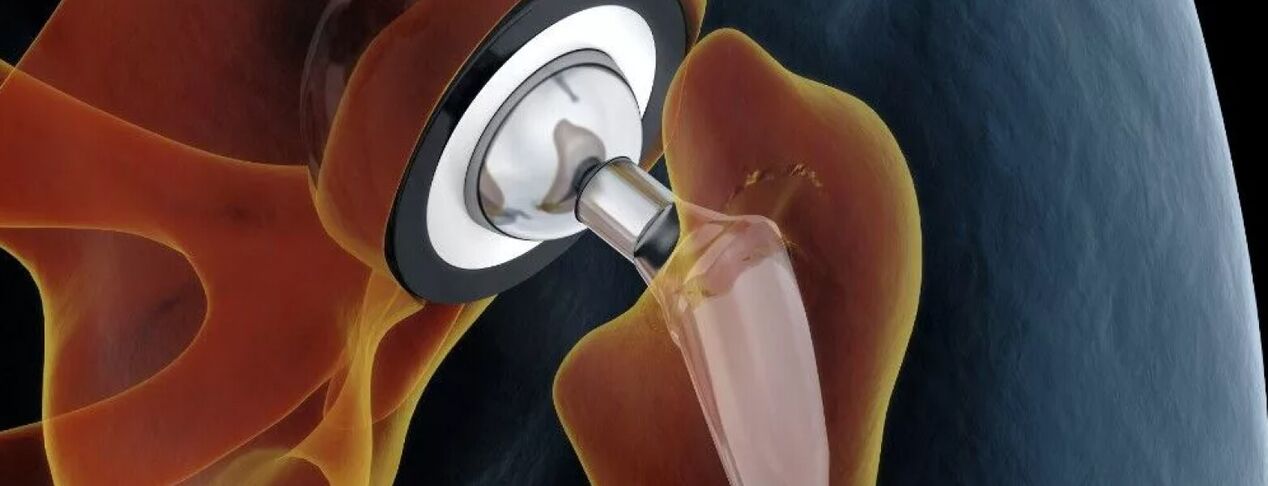
The gold standard for the treatment of severe coxarthrosis, including in the elderly, is hip arthroplasty. This operation involves replacing the destroyed hip joint with an artificial endoprosthesis made of durable and at the same time biologically compatible materials. Endoprosthetics allow you to fully restore the function of the hip joint, eliminate pain and return a person to a fully active life.
The essence of this type of surgical intervention is the resection of the femoral head and a small fragment of its neck. Also, the surgeon needs to prepare the surface of the acetabulum for the installation of the endoprosthesis, that is, remove all the osteophytes that have formed and achieve the maximum restoration of its normal shape. After that, the endoprosthesis of the chosen type is installed, which is fixed with special cement (preferably for the treatment of the elderly) or in a cementless way. In the second case, the endoprosthesis has a special spongy part that is in contact with the bone structure. Its fixation in the acetabulum is provided by the proliferation of bone tissue through the sponge.
For each patient, the type of arthroplasty is selected individually. The most effective is total arthroplasty, which involves the complete replacement of the entire hip joint, that is, the neck and head of the femur, as well as the acetabulum.
If the patient has preservation of normal hyaline cartilage on the surface of the acetabulum, he can undergo partial arthroplasty with replacement of only the femoral head and/or neck. For this purpose endoprostheses of different designs are used: monopolar and bipolar.
The only drawback of arthroplasty can be considered the need to replace the installed endoprosthesis after 15-30 years.
After endoprosthesis replacement, the patient is shown recovery, the duration of which depends on the rate of tissue repair. As part of rehabilitation, exercise therapy, physiotherapy and therapeutic massage are prescribed.
Before the advent of modern endoprostheses, patients with grade 3 coxarthrosis were prescribed osteotomy or arthrodesis. Today, this technique is used less and less, because it has several disadvantages. Therefore, arthrodesis involves fixing the bony structure of the hip joint with a metal plate. As a result, the pain syndrome is completely eliminated, but the joint loses its mobility completely. Therefore, after arthrodesis, the patient can only stand, but can no longer walk independently due to the lack of movement in the hip joint. Therefore, today arthrodesis is practically not carried out.
Osteotomy involves the implementation of an artificial fracture of the femur with a combination of bone fragments that will reduce the load on the affected hip joint. But the operation only gives a short-term effect, and in the future, the need for arthroplasty still arises.
Therefore, coxarthrosis of the hip joint is a rather dangerous disease, which can lead to disability. It seriously reduces the quality of life and takes away a person's work capacity. But if you pay attention to the early signs of pathology and seek advice from an orthopedic specialist in time, you can slow down its development and achieve a significant improvement in well-being. But with already running coxarthrosis, there is only one solution - arthroplasty. Fortunately, this method can be used even with serious degenerative-dystrophic changes and fully restore the normal function of the hip joint.